Resipher System
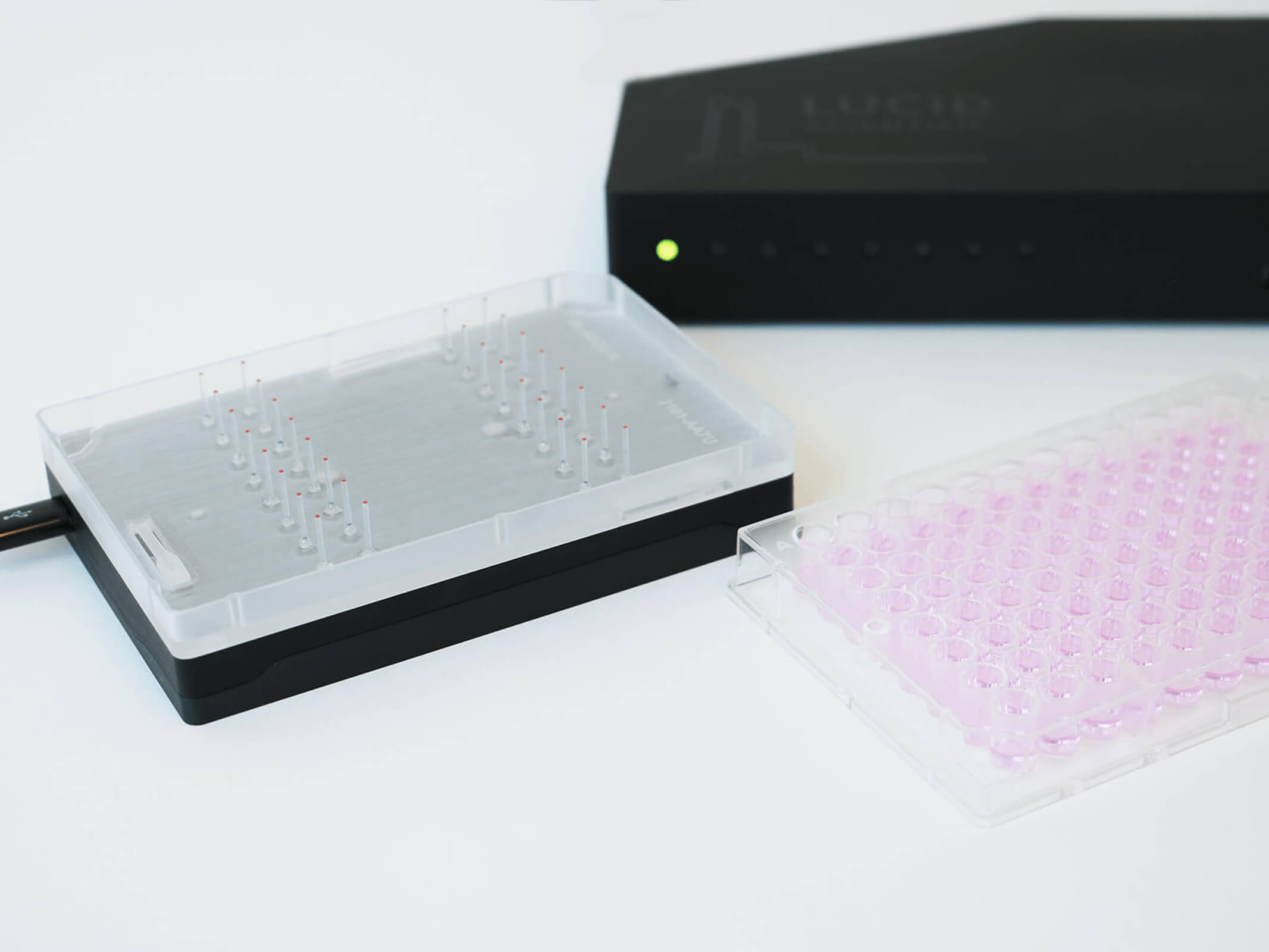
Turns a 96-well plate into a precise real-time oxygen consumption reader
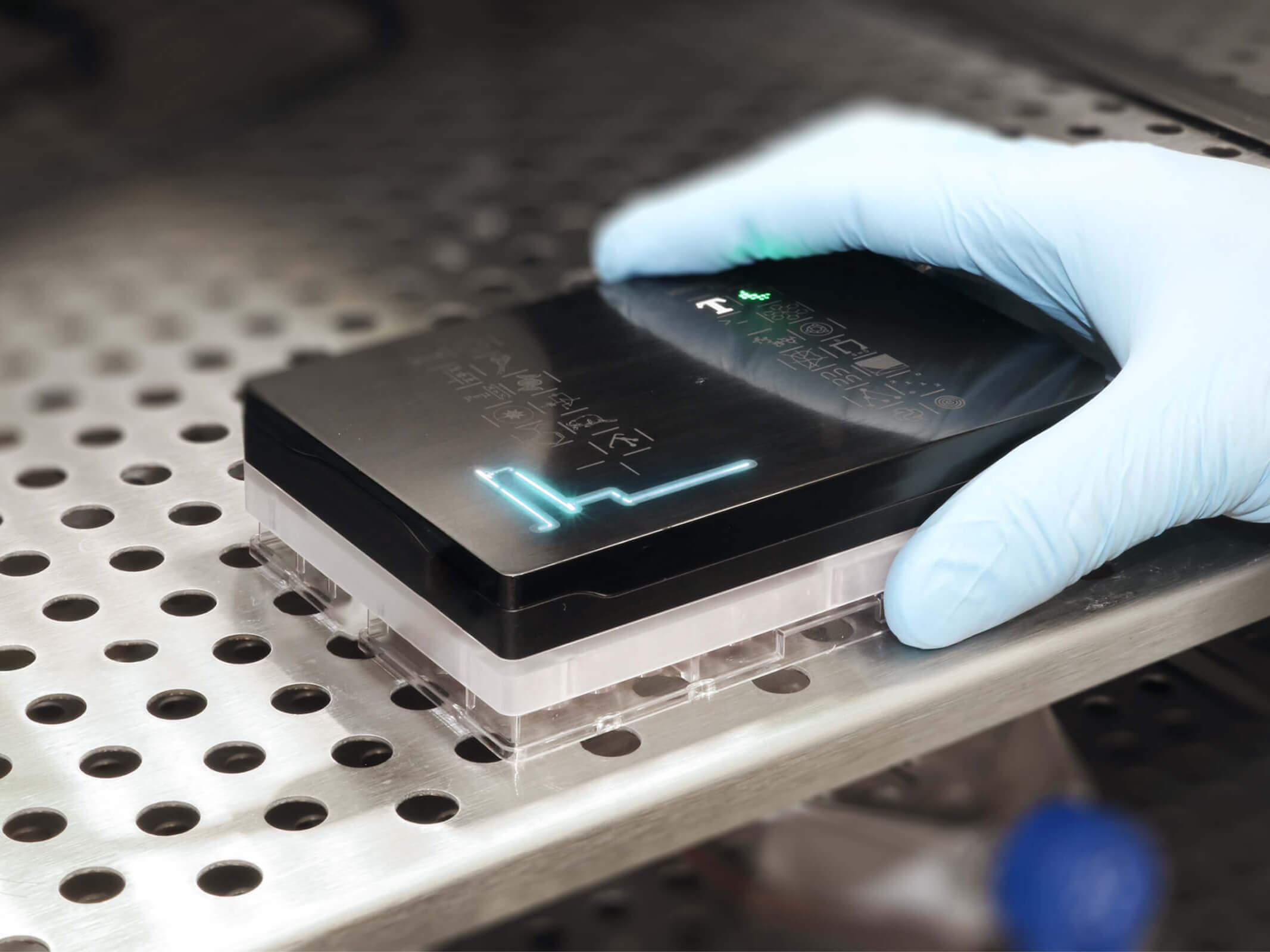
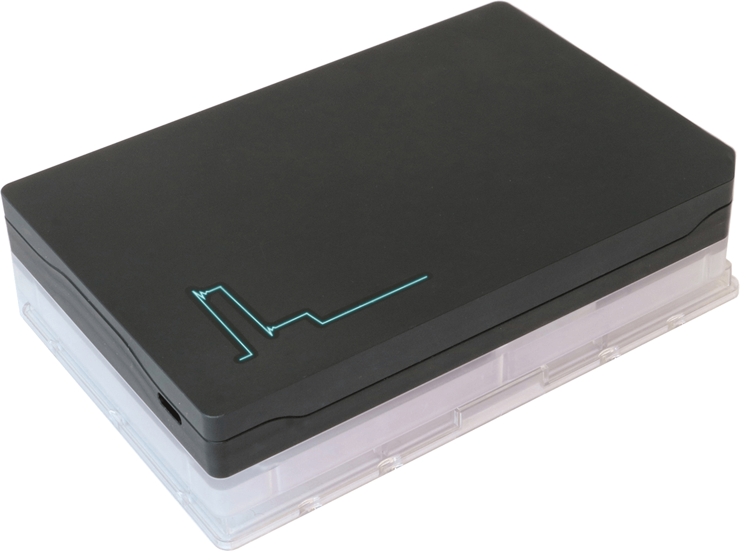
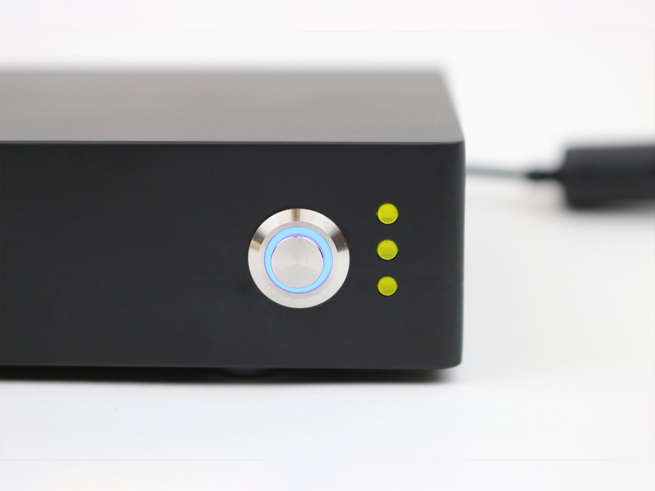
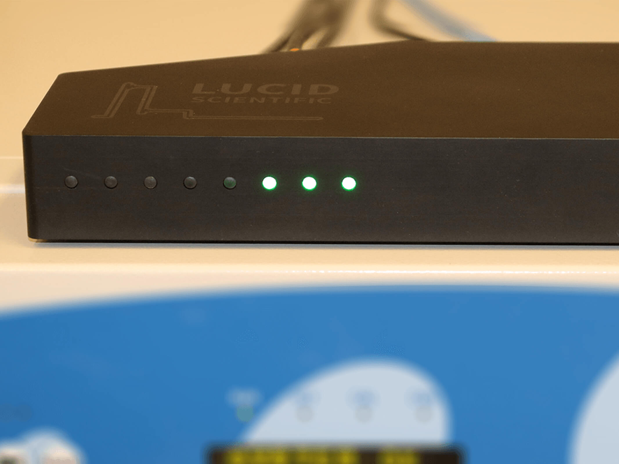
Lucid Scientific’s Resipher empowers researchers to precisely measure oxygen consumption directly in standard microplates. The system’s patented dynamic optical oxygen sensors provide the highest sensitivity without disturbing cells. Resipher’s super-compact profile sits directly in your incubator or workstation.
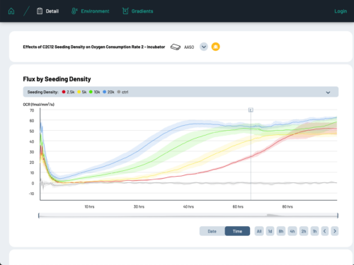
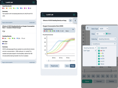
Resipher data is explored through Lucid Lab – a web-based, real-time logging and analysis software, providing fast and easy data visualisation for each well being monitored. Scientists can now watch their cellular experiments real-time from their computer or remotely via smartphone or tablet.
Real-time analysis
Resipher continuously monitors cell culture oxygen consumption inside the incubator. Data is streamed real-time to the cloud so you and your team can view on any connected device.
Use your current media
No need to reformat your workflow for analysis!
Innovative sensing
An array of ultra-sensitive dissolved oxygen sensors developed by Lucid performs dynamic vertical micro-movements that scan oxygen gradients formed by metabolically active cells.
Novel insights
Resipher provides researchers never-before-seen insights into dynamic cellular responses. High resolution gradient data for each well provides more in-depth analysis.
Cloud driven platform
Data is securely streamed through our cloud infrastructure as experiments happen. Our cross-platform app – Lucid Lab – enables your team to manage experiments, monitor performance, visualize data and share results from anywhere.
- Consumption resolution 5 fmol/mm2/sec
- Concentration sensitivity < 1µM
- Time resolution < 30 mins
- Resipher dimensions (mm) 128 (W) x 86 (D) x 15 (H)
- Run up to 8 devices from one hub
Lucid’s cross-platform web application allows teams to create and manage experiments, stream and monitor data from devices in real-time, and generate plots that are ready for publishing. Collaboratively record observations and events through the interface, share plots and experiments, and work from anywhere with full featured desktop and mobile versions.
Overview – https://youtu.be/QhpIty1lVP8
Intro Presentation – https://youtu.be/tX-pG9KiP0Q
Mobile demo – https://youtu.be/6vT8MofmJc0
Oxygen is a critical regulator of cellular metabolism and function in cell culture
Cell cycle induction in human cardiomyocytes is dependent on biosynthetic pathway activation
Considerations for using isolated cell systems to understand cardiac metabolism and biology
Fumarate is a terminal electron acceptor in the mammalian electron transport chain
- Algae
Amphidinium carterae, S major - Bacteria
E. coli - Human bone
SW1353, 143B - Human brain
U87 - Human brain endothelial
hCMEC/D3 - Human breast
MDA-MB-231, MCF7 - Human epithelial
HeLa - Human kidney
HEK 293 - Human large intestine/colon
HCT116, DLD1 - Human liver
HepG2 - Human lung
WI38, A549, CALU-3 - Human macrophage
KG-1, MV-4-11 - Human neurons
hESC - Human prostate
PC-3, InCAP, DU-145 - Human renal proximal tubule epithelial cells
RPTEC - Human retinal
H-RPE - Human stem
iPSC-RPE, E16 - Human T lymphocyte
Jurkat - Human umbilical vein endothelial cells
HUVEC - Monkey kidney
Vero - Mouse hippocampal
HT22 - Mouse macrophage
RAW 246.7 - Mouse muscle
C2C12 - Musculus (mouse) calvaria
MC3T3 - Rat muscle
L6 - Worm
C. Elegans - Other
Cardiomyocytes, Osteoblast, Osteoclast, Macrophage, Adipocyte, Fibroblast, Mesenchymal stem cells, iPSC – ECs, C16, Zebrafish embryos
Related products
-
Sage HLS
Extraction and Library Prep System
Price On Request £0.00 Add to basket -
Concept
Anaerobic workstation
Price On Request £0.00 Select options This product has multiple variants. The options may be chosen on the product page -
CondoCell
Transportable Hypoxia Containment vessel. Delivering luxury accommodation for cells!
Price On Request £0.00 Add to basket -
Concept 400 M
Micro-aerophilic workstation
Price On Request £0.00 Add to basket